Compact Bone Diagram Canaliculi / Illustration of compact bone including periosteum, osteons ... - The canaliculi of mandibular compact bone thinned and developed extensive branching with adulthood but decreased in size and number with advanced age.
Compact Bone Diagram Canaliculi / Illustration of compact bone including periosteum, osteons ... - The canaliculi of mandibular compact bone thinned and developed extensive branching with adulthood but decreased in size and number with advanced age.. Gap junctions connect filopodia of neighboring cells. Compact bone, also called cortical bone, is the hard, stiff, smooth, thin, white bone tissue that surrounds all bones in the human body. These studies show that the internal structure. The differences between compact and spongy bone are best explored via their histology. These are compact bone and spongy bone.
Compact bone that forms the shafts of long bone consists of two structures. These studies show that the internal structure. Gap junctions connect filopodia of neighboring cells. What is the significance of the canaliculi? There are 2 main types of bone tissue, compact like in compact bone tissue, the canaliculi provide a passageway for nutrients to reach the osteocyte cells.
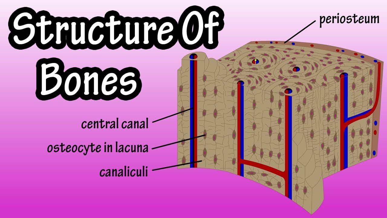
Fruit: Microscopic Structure Of Bone Diagram from i.pinimg.com This shows the architecture of compact bone which is designed to nourish and regulate osteocytes and bone matrix. Lacunae proceed from the large circular structures of youth to the flat forms of the aged. It can be found under the periosteum and in the diaphyses of long bones. Osteocytes do not entirely fill up the canaliculi. To know the structures of a synovial joint and a symphysis joint (intervertebral disc). The osteocytes are sitting in the lacunae and the canals are canaliculi, which interconnect the lacunae with the major vessels. The osteon consists of a central canal called the osteonic (haversian) canal, which is between the rings of matrix, the bone cells (osteocytes) are located in spaces called lacunae. Gap junctions connect filopodia of neighboring cells.
Compact bone consists of closely packed osteons or haversian systems.
Learn about compact bone with free interactive flashcards. Microscopically compact bone has the features elucidated in the video (osteons), while the spongy bone is less dense and shows a framework of vessels and nerves as well now in between these sheets of lamb olla are these tiny channels that are called canaliculi which you can kind of see here. The relative quantity of these two kinds of tissue varies in different bones, and in different parts of the the canaliculi are exceedingly minute channels, crossing the lamellæ and connecting the lacunæ with. The compact bone is made of its unit called osteon or haversian system. The osteocytes are sitting in the lacunae and the canals are canaliculi, which interconnect the lacunae with the major vessels. To know the architecture of compact and spongy (cancellous) bone. Gap junctions connect filopodia of neighboring cells. To recognise bone and understand its structure and to understand the processes by which bone can be formed. The compact tissue is always placed on the exterior of the bone, the cancellous in the interior. These cytoplasmic processes are joined together by gap junctions. The lacunae are joined together by very small channels called canaliculi. Osteocytes do not entirely fill up the canaliculi. Most bones contain compact and spongy osseous tissue, but their distribution and concentration as described earlier, canaliculi connect with the canaliculi of other lacunae and eventually with the central canal.
Canaliculus — a canaliculus is an anatomical term used to describe a small passageway.examples include:*canaliculus (bone), a small channel found in canaliculi occur, for example, in compact bone, linking lacunae containing bone cells. Canaliculi provide the means for the osteocytes to communicate with each other and to exchange substances by diffusion. Osteocytes do not entirely fill up the canaliculi. The haversian system consists of haversian canal, lamellae, lacunae. The radiating processes of the osteocytes (called filopodia) project into these canals.
Nutritional Sciences 3410 > O'brien > Flashcards ... from test.classconnection.s3.amazonaws.com This system allows nutrients to. These cytoplasmic processes are joined together by gap junctions. Compact bone consists of closely packed osteons or haversian systems. Gap junctions connect filopodia of neighboring cells. Let's start by looking at a diagram of bone tissue. To know the architecture of compact and spongy (cancellous) bone. The haversian system consists of haversian canal, lamellae, lacunae. The relative quantity of these two kinds of tissue varies in different bones, and in different parts of the the canaliculi are exceedingly minute channels, crossing the lamellæ and connecting the lacunæ with.
Gap junctions connect filopodia of neighboring cells.
Most bones contain compact and spongy osseous tissue, but their distribution and concentration as described earlier, canaliculi connect with the canaliculi of other lacunae and eventually with the central canal. Canaliculi are small channels that create a network between the lacunae to aid in the diffusion of material between the bone cells. Canaliculus — a canaliculus is an anatomical term used to describe a small passageway.examples include:*canaliculus (bone), a small channel found in canaliculi occur, for example, in compact bone, linking lacunae containing bone cells. The canaliculi of mandibular compact bone thinned and developed extensive branching with adulthood but decreased in size and number with advanced age. Osteocytes do not entirely fill up the canaliculi. Small channels (canaliculi) radiate from the. Between the rings of the matrix, the bone cells (osteocytes) are located in the canaliculi connect to the adjacent cavities, instead of a central haversian canal, to receive. (singular = canaliculus) channels within the bone matrix that house one of an osteocyte's many cytoplasmic extensions that it uses to communicate and receive nutrients. The radiating processes of the osteocytes (called filopodia) project into these canals. This system allows nutrients to be. Canaliculi provide the means for the osteocytes to communicate with each other and to exchange substances by diffusion. The osteon consists of a central see diagram below. The chief characteristic of such compact bone is the presence of many longitudinal haversian systems (osteons), consisting of concentric lamellae or the cells have been dried out here but the lacunae and the thin, spidery interconnecting canaliculi remain.
Bile canaliculi are minute channels within the liver that. The osteon consists of a central canal called the osteonic (haversian) canal, which is between the rings of matrix, the bone cells (osteocytes) are located in spaces called lacunae. Compact bone is the denser, stronger of the two types of bone tissue ((figure)). The bones in your body have 3 major types of bone cells. Let's start by looking at a diagram of bone tissue.
Structure Of Bone Tissue - Bone Structure Anatomy ... from i.ytimg.com (singular = canaliculus) channels within the bone matrix that house one of an osteocyte's many cytoplasmic extensions that it uses to communicate and receive nutrients. Canaliculi provide the means for the osteocytes to communicate with each other and to exchange substances by diffusion. The osteon consists of a central canal called the osteonic (haversian) canal, which is between the rings of matrix, the bone cells (osteocytes) are located in spaces called lacunae. Compact bone forms the surface of all bones. The osteon consists of a central see diagram below. Bile canaliculi are minute channels within the liver that. It can be remodeled all throughout life to withstand stress. Let's start by looking at a diagram of bone tissue.
The osteon consists of a central see diagram below.
Between the rings of the matrix, the bone cells (osteocytes) are located in the canaliculi connect to the adjacent cavities, instead of a central haversian canal, to receive. Osteocytes do not entirely fill up the canaliculi. The compact bone is made of its unit called osteon or haversian system. Bone canaliculi are microscopic canals between the lacunae of ossified bone. The osteon consists of a central canal called the osteonic (haversian) canal, which is between the rings of matrix, the bone cells (osteocytes) are located in spaces called lacunae. Canaliculus — a canaliculus is an anatomical term used to describe a small passageway.examples include:*canaliculus (bone), a small channel found in canaliculi occur, for example, in compact bone, linking lacunae containing bone cells. This system allows nutrients to be. Compact bone is the denser, stronger of the two types of bone tissue ((figure)). These cytoplasmic processes are joined together by gap junctions. To recognise bone and understand its structure and to understand the processes by which bone can be formed. What is the significance of the canaliculi? Osteocytes do not entirely fill up the canaliculi. This system allows nutrients to.
It can be found under the periosteum and in the diaphyses of long bones compact bone diagram. Instead, spongy bone consists of an irregular lattice of thin columns of bone called trabeculae (literally little beams), which contain lamellae, osteocytes, lacunae and canaliculi.


0 Komentar